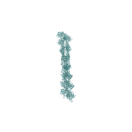
Human Reproduction: Fertilisation to Implantation
- Biology
- Class 10–12
- 3D model
- English
Human Reproduction: Fertilisation to Implantation: a 3D model for Class 10–12 Biology, covering the ovaries release oocytes (egg cells); the testes make sperm cells, sperm are stored and mature in the epididymis and are carried by the vas deferens and urethra, seminal vesicles, prostate and bulbourethral glands add fluids; these secretions are not sperm cells, the fimbriae guide the released oocyte into the oviduct, fertilisation normally happens in the oviduct near the ampullary-isthmic junction, not in the uterus or ovary.
The 3D viewer and AR open with a free Lably account.
Also called
human reproduction · reproductive system · female reproductive system · male reproductive system · fertilisation · fertilization · implantation · zygote · gametes · sperm and egg · ovulation · oviduct · fallopian tube · uterus · blastocyst · cleavage · morula · how do organisms reproduce
Topics
About this model
A GLB file of about 1514 KB, shown in Lably's 3D viewer and, on a phone, placed in your room with AR. Credit: Uterus and ovary: HuBMAP Human Reference Atlas, CCF 3D Reference Object Library (Kristen Browne et al., NIH/NIAID; derived from the NLM Visible Human Project), humanatlas.io, CC BY 4.0 - uterus voxel-remeshed, turned upright, cut open and split into wall, lining and cervix; ovary also mirrored for the right side. Zona pellucida, oocyte cytoplasm and sperm head/midpiece: 'Human egg cell' and 'Sperm cell' by Julio Hidalgo, Tactiles (tactiles.eu), CC BY-SA 4.0 - decimated, re-centred, print keys flattened; sperm tail replaced by an authored rig. Male organ schematic, oviducts, fimbriae, vagina, nuclei and chromosomes, cleavage stages, blastocyst, markers and all animation newly authored for Lably. The whole model is shared under CC BY-SA 4.0. | CC BY-SA 4.0